News Center
Some knowledge points of DR system, pet hospital DR instrument manufacturer tells you
Classification:
Release time:
2022-08-17
Pet hospital DR instrument productionManufacturers believe that in recent years, with the rapid development of radiological imaging technology, digital X-ray photography system in clinical application is more and more common. DR refers to a new technology of digital X-ray photography under the direct control of a computer, that is, the use of amorphous silicon flat-panel detectors to convert the X-ray information penetrating the human body into digital signals, and the computer reconstructs the image and performs a series of image post-processing. Due to the use of digital technology, various image post-processing can be performed according to clinical needs.
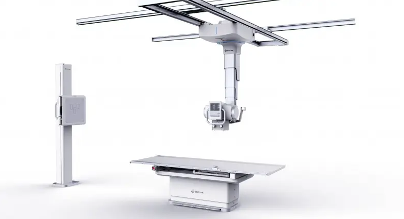
Pet hospital DR instrument productionManufacturers believe that DR is a digital imaging system. Medical DR refers to the hospital radiology department to shoot the film. On the basis of traditional X-ray, DR equipment increases the digital process of computer processing, so that the image quality is higher, the time is shorter, and the amount of radiation is smaller.
Pet hospital DR instrument productionAccording to the X-ray exposure method, the DR system can be divided into surface exposure imaging technology and line scanning imaging technology. The main difference between the two techniques is the detector acquisition method.
1. Surface exposure imaging technology: The main feature is that the detector uses a large area array detector design, also called a flat panel detector. Another feature is that at the moment of X-ray exposure, the basic information of the imaging area of the examined human body can be collected at one time. At present, amorphous silicon, amorphous selenium, CCD and other flat panel detectors are used in this way.
2. Line scanning imaging technology: pet hospital DR instrument manufacturers believe that when exposed, X-ray irradiation field is fan-shaped perpendicular to the human body, scanning the human body examination area at a constant speed along the long axis of the human body. At present, the main ways to use this method are: multi-wire proportional ionization chamber gas detector; scintillation crystal/photodiode linear array detector: solid-state semiconductor/CMOS linear array detector.
Pet hospital DR instrument manufacturers believe that the commonly used DR classification is based on the energy conversion mode of the X-ray detector. The energy conversion mode of the X-ray detector includes a direct conversion mode and an indirect conversion mode:
1. Direct conversion: direct digital X-ray photography. The pet hospital DR instrument manufacturer believes that the basic principle is to project X-rays on the X-ray detector, and the photoconductive semiconductor material collects X-ray photons and converts the X-ray intensity distribution into electrical signals. Currently commonly used photoconductive semiconductor materials are amorphous selenium (a-Se), lead iodide (PbI2), mercury iodide (HgI) and so on. X-ray detectors that have been used in DR equipment are mainly amorphous selenium flat panel detectors and cadmium telluride/cadmium zinc telluride linear detectors.
2. Indirect conversion: pet hospital DR instrument manufacturers believe that compared with the direct conversion method, indirect digital X-ray photography is to project X-rays on the X-ray detector, which first irradiates some scintillating crystal material. After the crystal absorbs the amount of X-rays, it releases energy in the form of visible fluorescence, transmits through the space circuit, is collected by the photodiode, and is converted into an electrical signal. Luminescent crystalline substances for indirect conversion mainly include cesium iodide (CsI) and gadolinium oxysulfide (GOS).
News
Related News
Electronic and Electrical Industrial Park, Zhengzhou High-tech Industrial Development Zone

WeChat Public